Saturday, May 12, 2012

Rilpivirine: A New HIV/AIDS Treatment


3D Modeling of the Human Immunodeficiency Virus [1]

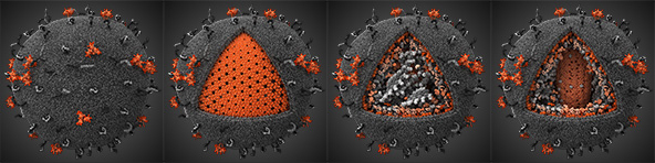
Growing up in the 1990’s and being the son of an infectious disease physician, I had always been warned of the dangers of HIV. Particularly, I was taught about the fact that there is no cure once one is infected with the virus. As I grew up and became more educated in the biological sciences, I became curious to why we essentially are able to prevent certain viral infections (such as the chicken pox virus) and not have any way to prevent other viral infections such as HIV. “Aren’t all viruses the same and why can’t we ever truly be cured of a viral infection?” The short answer, which I would come to learn, is that there is a large amount of diversity among the different viruses that can infect our bodies and hijack our essential life systems. However, a universal key mechanism of viral infections that makes them extremely hard to cure is that there is incorporation of viral genetic material into our own genome of an affected cell; essentially making the virus a part of us. Click the image below to explore the replication (life cycle) of the HIV virus.


HIV Replication Cycle [2]
My Dad and I at dinner


So, when I came across the article titled Experimental vaccine helps protect monkeys against AIDS-like infection on CNN Health a month back I was immediately intrigued. The article stated that the developed vaccine had been successful in treating SIV (Simian Immunodeficiency Virus), a close relative of the human HIV virus and the primary animal model virus for HIV. Although the vaccine had been successful in initially preventing clinical SIV infection in the tested monkeys, each individual monkey eventually developed SIV after repeated exposures. Despite the vaccines eventual failure to completely ameliorate the potential for SIV infection, the scientific community believes that they are now a step closer to finding a preventative cure for HIV. After reading the article and realizing that HIV has not been cured, I became curious about what kinds of treatments there are on the market to treat someone with HIV/AIDS. 

I decided to preform a basic google search for “HIV/AIDS Treatments.” The search resulted with the the Mayo Clinic website article outlining the different treatments. The article stated that there are a wide variety of drug treatment options. These treatments included Non-nucleoside reverse transcriptase inhibitors (NNRTIs), nucleoside reverse transcriptase inhibitors (NRTIs), protease inhibitors (PIs), entry of fusion inhibitors, and integrase inhibitors. With many kinds of treatments to chose from, I discovered that treatment in the clinic for HIV/AIDS involves a cocktail of different treatment types. Fascinated by HIV’s ability to convert its RNA genetic code into DNA, I dug further to investigate how the different drugs on the market affect reverse transcriptase, the enzyme that is responsible to convert RNA to DNA.

The two classes of drugs that affect reverse transcriptase action are NNRTIs and NRTIs. NNRTIs act by antagonistically binding to the enzyme and halting its function where NRTIs utilize faulty versions of the building blocks that reverse transcriptase uses to convert RNA to DNA. This essentially prevents a good DNA copy of the viral genome to be produced. A key difference between NRTIs and NNRTIs is that one is a competitive inhibitor while the other is a non-competitive inhibitor of the enzymatic process. NRTI’s are competitive inhibitors. They compete with the cells normal nucleosides (DNA building blocks) for integration into the viral DNA strand. On the other hand NNRTIs are non-competitive inhibitors meaning that they are a particular kind of molecule that reduces enzyme activity by binding to a site that is different than the active site of a molecule where it does not face binding competition [3]. 
Crystallographic Structure of Reverse Transcriptase where the P51 subunit is colored green
and the P66 subunit is colored cyan [4]

After another quick Goggle search for NNRTIs, I came to learn that the FDA had recently approved a new drug called Rilpivirine (trade name Edurant) in May 2011. I became instantly curious to learn the novel mechanism of action by which this drug inhibits reverse transcriptase. Like all NNRTI’s, Rilpvirine (see Marvin Sketch Below) inhibits reverse transcriptase by allosteric binding. This type of inhibition works by non-competitively binding to reverse transcriptase at a location other than the enzyme’s active site, resulting in inhibition of its function. For Reverse Transcriptase this binding site is known as the Non-Nucleoside Inhibitor Binding Pocket (NNIBP). But what about this drug makes it unique?


Figure 2. Mouseover sends to MarvinSketch.
Before I searched for the answer to this question, I sought to understand the structure of reverse transcriptase, specifically the NNIBP, as well as the characteristics that make a good HIV/AIDS drug. A preformed a quick Google search resulted for "reverse transcriptase" and ended up finding ample information. The first link that I clicked on was the Wikipedia page for Reverse transcriptase. From the wiki page I learned that Reverse transcriptase is a type of polymerase that is RNA-dependent DNA synthesizing and that it is made up of two distinct components, the p51 and p66 subunits. The p66 subunit contains the fingers, palm and thumb of the enzyme and the p51 subunit contains an additional set of fingers. The analogy of reverse transcriptase as a hand is used since it describes which parts of the enzyme are squeezing together to connect the different nucleosides in a chain. Use the Jmol applet below to explore the structure of Reverse Transcriptase. 

The Non-nucleoside inhibitor-binding pocket (NNIBP) is located on the P66 subunit. This pocket has a characteristic positive charge and hydrophobicity due to the presence of particular amino acids including Val 106, Tyr 188, Trp 229, Leu 234, Leu 100, Phe 227, Tyr 319, Pro 236, Tyr 181, Lys 103, Gly 190. During protein synthesis these amino acids come together to form a pocket (via hydrophobic interactions) to shield themselves from the highly polar hydrophilic intracellular space. The NNIBP is located approximately 10 angstroms away from reverse transcriptase’s active binding site. The active binding site is the location where reverse transcriptase converts the viral RNA into DNA. NNRTIs target the NNIBP and causing a change in steric interactions that results in inactivation of reverse transcriptase’s function. 

I found the answer to my second question "what makes a good anti-HIV/AIDs medication?" in an article published in the Journal of Medicinal Chemistry. The article stated that a good anti-HIV drug should have the following properties: (1) it should be highly active against wild-type and mutant types of HIV without allowing breakthrough, (2) it should have high oral bioavailability and long elimination half-life, allowing for once-daily oral treatment at low doses, (3) have minimal adverse effects, and (4) be easy to synthesize and formulate [5]. With a brief and basic understanding of the structure and properties of Reverse transcriptase and its inhibition binding pocket as well as the key properties of a well engineered anti-HIV drug, I was able to move forward and learn more about Rilpivirine. Specifically, how it acts as a NNRTI as well as how it is different from other NNRTIs that are available on the market. 

Rilpivirine is a diarylpyrimidine and has the structural formula 4-[[4-[[4-[(E)-2-cyanoethenyl]-2,6-dimethylphenyl]amino]2-pyrimidinyl]amino]benzonitrile. In lay terms, Rilpivirine is made up of three cyclic rings, two nitrile groups, and a conjugated double bond system. Like most diarylpyrimidine non-nucleoside reverse transcriptase inhibitors, I learned that Rilpivirine has characteristic flexibility that gives it the ability to maintain its activity in situations where wild-type reverse transcriptase is not present. Also, Rilpivirine’s targets certain amino acids which display low levels of mutability which helps give the drug very low resistance against mutant reverse transcriptase’s. Explore Rilpivirine's structure using the Jmol applet below.

It is Rilpivirine’s ability to interact with both mutant and wild type Reverse Transcriptase that makes it a novel NNRTI. Originally, NNRTI’s were not effective in treating patients when mutant Reverse Transcriptase was present. The resistance towards older NNRTI's was due impart to decreased torsion and flexibility among the drugs chemical bonds. Another key feature of early NNRTIs was their dependence on π -stacking interaction with specific Tyrosine groups (Y181 and Y188). Evidently, it turned out that these specific Tyrosine groups exhibit high mutation rates and accounted for the most common type of observed mutant reverse transcriptase in the clinic. Since the binding mechanism of these NNRTI’s relied on highly stable and ridged π-stacking interactions, when these amino acid groups mutated, the drugs had no way of conforming to fit the new mutant NNIBP. Rilpivirne, on the other hand, relies on a slightly different mechanism of binding and employs greater torisonal flexibility. The torsion angle diagram shows the degree of rotation around certain sigma bonds when Rilpivirine is bound to its binding pocket. Despite the single degree of rotational difference between the wild-type and mutant version of Reverse Transcriptase, researchers believe it is this extra degree that makes all the difference. 


Rilpivirine Torsion Degrees in Wild-Type & Mutant Reverse Transcriptase [6]


Instead of targeting Y181 and Y188, Rilpivirine targets a specific Tryptophan group (W299). In Reverse Transcriptase mutant analysis, it has been shown that this specific Tryptophan group shows stability, with regards to mutability, across all mutants making it the perfect target for a NNRTI with low levels of drug resistance. As shown in the figure below, Rilpivirine’s nitrile groups give it the ability to specifically target and interact with Tryptophan 229 and Tyrosine 318. These highly polar functional groups are able to interact via dipole-dipole interactions with W229 and Y318 helping to Rilpivirine to settle into the binding pocket. With regards to W229, the nitrile group interacts via an edge to face interaction. It is also possible that these nitrile groups could be undergoing H-bonding even though they are poor hydrogen acceptors. Like previous NNRTIs, Rilpivirine relies on π-stacking, hydrophobic interactions as well as electron rich and electron deficient interactions.
Rilpivirine in the NNIBP [5]
Although mechanistically the theory behind how Rilpivirine would interact with Reverse Transcriptase and inhibit its function makes sense, it is often found that in vivo, drugs and treatments have very unpredictable actions. With all drug development there is the inherent risk that the drug you developed to treat a certain disease will cause harm in another part of the body. Since Rilpivirine was approved by the FDA, it is safe to say that it is a safe and effective drug to treat HIV. However, how does Rilpivirine compare to other drugs that have been proven to be successful NNRTI drug treatments. A quick search on the FDA’s website returned the clinical data from its stage three clinical drug trials in which  a comparison study between Rilpivirine and the leading drug on the market, Efavirenz, was conducted. See graphs below that compare Rilpivirine and Efavirenz effectiveness by comparing measured CD4 cell counts and viral RNA concentration of treated patients. 


Percentage of patients with a viral load of less than 50 copies per mL from baseline to week 48 (A) and mean change in absolute CD4 cell count from baseline (B)(A) Intention-to-treat time-to-loss-of-virological-response algorithm. (B) At week 48, mean change in absolute CD4 cell count from baseline was 196 cells per μL (95% CI 179–212) for rilpivirine and 182 cells per μL (165–198) for efavirenz (p=0·13). [8]

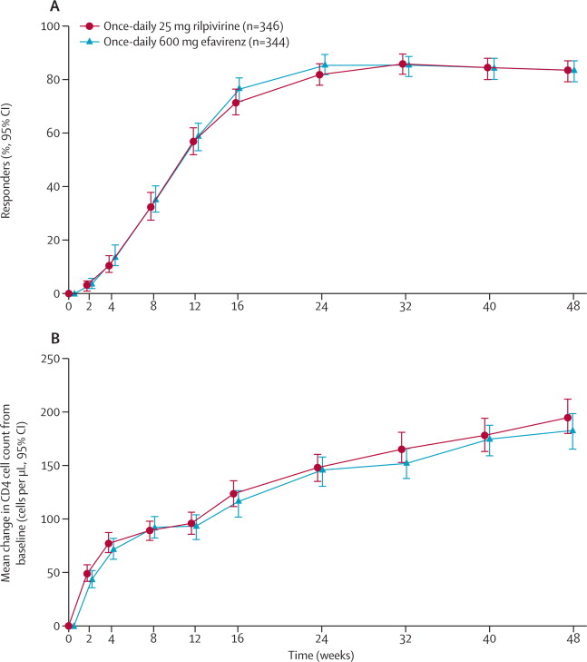
The data shows that Rilpivirine is an equally effective drug in comparison to Efavirenz. From the graphs above both CD4 cell count (graph B) and viral load (graph A) for patients treated with Rilpivirine showed similar disease efficacy. Over time, CD4 cell counts increased and Viral Load plateaued in treated patients. Despite the similar clinical results in patients treated with either Rilpivirine or Efavirenz, it is important to remember that Rilpivirine is overall a better drug of choice. It shows low levels of resistance to differing mutant forms of Reverse Transcriptase, has been shown to have fewer side effects while still meeting the effectiveness of previous drugs on the market. Rilpivirine in conjuction with other HIV treatments should help to improve the lives of living with this disease. Although, the cure for HIV/AIDS has not been discovered it is these small advances in medical treatment and research that help to pave the way for hope and aspiration that one day a cure of HIV will be discovered. 

References:

[1] "Human Immunodeficiency Virus Model." Human Immunodeficiency Virus Model. Ed. Ivan Konstantinov. Visual Science, 8 Sept. 2010. Web. 12 May 2012 <http://visualscience.ru/en/projects/hiv/illustrations/>.

[2] National Institute of Allergy and Infectious Disease. "HIV Replication Cycle." HIV Replication Cycle. National Institute of Health, 3 Apr. 2012. Web. 12 May 2012. <http://www.niaid.nih.gov/topics/HIVAIDS/Understanding/Biology/pages/hivreplicationcycle.aspx>. 

[3] National Institues of Health Chemical Genomics Center. "Types of Inhibition." - Assay Guidance Wiki. National Institues of Health Chemical Genomics Center, 20 Sept. 2010. Web. 12 May 2012. <http://assay.nih.gov/assay/index.php/Types_of_Inhibition>. 

[4] "RCSB Protein Data Bank - RCSB PDB - 1HMV Structure Summary." RCSB Protein Data Bank - RCSB PDB - 1HMV Structure Summary. RCSB Protein Data Bank. Web. 12 May 2012. <http://www.rcsb.org/pdb/explore/explore.do?structureId=1hmv>.

[5] Janssen, Paul A. J., Paul J. Lewi, Eddy Arnold, Frits Daeyaert, Marc De Jonge, Jan Heeres, Luc Koymans, Maarten Vinkers, Jérôme Guillemont, Elisabeth Pasquier, Mike Kukla, Don Ludovici, Koen Andries, Marie-Pierre De Béthune, Rudi Pauwels, Kalyan Das, Art D. Clark,, Yulia Volovik Frenkel, Stephen H. Hughes, Bart Medaer, Fons De Knaep, Hilde Bohets, Fred De Clerck, Ann Lampo, Peter Williams, and Paul Stoffels. "In Search of a Novel Anti-HIV Drug:  Multidisciplinary Coordination in the Discovery of 4-[[4-[[4-[(1)-2-Cyanoethenyl]-2,6-dimethylphenyl]amino]-2- Pyrimidinyl]amino]benzonitrile (R278474, Rilpivirine)." Journal of Medicinal Chemistry 48.6 (2005): 1901-909. Print.

[6] Lansdon, Eric B., Katherine M. Brendza, Magdeleine Hung, Ruth Wang, Susmith Mukund, Debi Jin, Gabriel Birkus, Nilima Kutty, and Xiaohong Liu. "Crystal Structures of HIV-1 Reverse Transcriptase with Etravirine (TMC125) and Rilpivirine (TMC278): Implications for Drug Design." Journal of Medicinal Chemistry 53.10 (2010): 4295-299. Print.

[8] Rilpivirine versus efavirenz with tenofovir and emtricitabine in treatment-naive adults infected with 

HIV-1 (ECHO): a phase 3 randomised double-blind active-controlled trial; Dr, Prof Jean-Michel 

Molina MD,Pedro Cahn MD,Beatriz Grinsztejn MD,Prof Adriano Lazzarin MD,Anthony Mills 

MD,Prof Michael Saag MD,Prof Khuanchai Supparatpinyo MD,Prof Sharon Walmsley MD,Herta 

Crauwels PhD,Laurence T Rimsky PhD,Simon Vanveggel MSc,Katia Boven MD,on behalf of the 

ECHO study group The Lancet - 16 July 2011 ( Vol. 378, Issue 9787, Pages 238-246 ) DOI: 

10.1016/S0140-6736(11)60936-7

Friday, May 4, 2012


Adderall and Performance Enhancement in Students
Gayatri Satam

Adderall is an amphetamine used most commonly as a medication for attention deficit hyperactivity disorder and narcolepsy. It works by directly targeting dopamine in the mesolimbic reward pathway. Adderall works by binding to monoamine transporters in the pathway and thus increasing levels of dopamine, norepinephrine, and serotonin in the system. Adderall works not only by increasing the release of more neurotransmitters into the synapse, but also by delaying their time in the synapse by slowing down their removal. Especially in a college setting, Adderall seems to be widely used as a performance enhancing drug used to increase concentration. According to a study in 2005 in the journal Addiction, about 25% of students on college campuses (both undergraduate and graduate) use non-prescribed stimulants to enhance function. My preliminary research made me wonder, how is a drug like Adderall used by students to enhance their learning and studying abilities? Furthermore, how does it work in the brain to elicit this high performance outcome?

Amphetamines like Adderall are primarily releasers of dopamine, norepinephrine, and small amounts of serotonin. At higher doses, they have also been seen to inhibit the reuptake of dopamine and norepinephrine, but not to the levels of cocaine. Dopamine has been thought to play an important role in memory formation whereas norepinephrine is linked with increased attentiveness and arousal. Amphetamines have been seen to be associated with feelings of alertness, elevation of mood, drive, ability to concentrate, and euphoria. The first step in the mechanism of action of amphetamines like Adderall is the rate limitation of tyrosine hydroxylase to convert 1-tyrosine into 1-dopa, which begins the production of dopamine, norepinephrine, and epinephrine. Amphetamine also works to inhibit the enzymes monoamine oxidase A and B in substantially high doses. These enzymes are primarily used for the breakdown of neurotransmitters like serotonin, dopamine, norepinephrine, and epinephrine. This results in the accumulation of neurochemicals in the synapse.

Figure 1. Below is an image depicting Adderall binding to and blocking monoamine transporters in the neuronal synapse. Monoamine transporters function in the reuptake of neurotransmitters from the synapse.


Figure 2. This is a picture of the structure of Adderall.


The mesolimbic pathway that Adderall is involved with is strongly linked to feelings of reward and desire and thus has been shown to be involved in the onset of disorders like depression, addiction, and schizophrenia. The mesolimbic pathway begins in region knokn wn as the ventral tegmental area of the midbrain and connects to the medial prefrontal cortex and the limbic system (shown below).


Studies have shown that mice that have had their ventral tegmental areas removed have not shown a loss in learning abilities, but rather a loss in the motivation to work for any particular reward. Dopamine, which is the main candidate involved in the mesolimbic pathway, has thus been heavily studied in its effect on attention. It has been understood that a lack of dopamine stimulation has been a key factor in disorders like ADHD and ADD. Adderall works to relieve this problem by stimulating dopamine and also by releasing dopamine reuptake inhibitors.

Figure 3. The life cycle of a normal neurotransmitter.

Figure 4. The structural similarity between Adderall and Dopamine (highlighted in blue). It shows how Adderall can be used to bind to the same receptors as dopamine.

Based off of these findings, I would speculate that increasing the levels of dopamine in a regular student would similarly work to improve their attention on a particular topic. As of this far, I would think that Adderall works to increase dopamine (or decrease the uptake of dopamine) which thereby increases the motivation that students need to complete the task at hand. The problem with drugs like Adderall is that they can become habit forming and can lead to severe consequences like heart problems, sleep deprivation, and most commonly, addiction. Amphetamines are classified as Class II medications, which means that they can have the potential to become addictive and abused as drugs. This is because the brain is tricked into thinking that it does not need dopamine anymore. When the reuptake receptors are blocked, the dopamine cannot become recycled and reused therefore the overall concentration of dopamine decreases causing future problems for abusers.

In my quest to understand the mechanism of Adderall and its effect on concentration and motivation of a student, I also came across two other drugs that were often mentioned: Ritalin (methylphenidate) and Dexedrine (dextroamphetamine). All three of these drugs are used to relieve the symptoms of ADHD and ADD, yet I was unsure of the differences between them, and if these drugs could also be used in the same way as Adderall is. The first thing I found out was that Dexedrine was an amphetamine, while Ritalin was not, meaning that it doesn’t affect the same pathway as Adderall and Dexedrine do. Dexedrine is also composed of one amphetamine salt whereas Adderall is composed of four. This means that a much higher dosage of Dexedrine is required to elicit the same response as Adderall. However, due to the higher number of amphetamine salts in Adderall, it has also been shown to result in more side-effects than does Dexedrine. These side effects include increased heart rate, blood pressure, inability to fall asleep, and decreased appetite. Due to these effects, Dexedrine is found to be substantially safer and thus is more widely sold unlike Adderall which is only legal in the United States and Canada. Ritalin, unlike both Adderall and Dexedrine, has the least potential for addiction.

The three drugs that I came across most: Adderall, Ritalin, and Dexedrine have each been shown to have a direct effect in performance enhancement, particularly in the boost of concentration and motivation that is necessary for long hours of studying and cramming. Overall, the pathways of all three of these drugs target the same thing: dopamine levels. In general, they work to increase levels of dopamine and also increase the amount of time dopamine spends in the synapse between two neural cells. Dopamine works by increasing alertness, thereby allowing a student to focus better on their work.

References:
The drawn image was based off a figure found from http://www.ncbi.nlm.nih.gov/books/NBK11110/figure/A386/?report=objectonly
The image of Adderall in the neuronal synapse was taken from: http://www-scf.usc.edu/~uscience/adderall_abuse.html


Protein Supplementation in Athletes


As a Kinesiology major, I know many active people that are concerned with their health and are aware that the overall quality of health of an individual can be improved by engaging in physical activity and regular exercise. One of the popular topics of conversation and something that is seen on a regular basis in advertisements is the use of protein supplements, which are supposedly helpful in keeping an athlete active and strong throughout tough workout regimens. Personally, I was first exposed to the idea of an increased protein diet from my younger brother, who is a high school athlete that engages in a high amount of physical activity on a daily basis. At the young age of fifteen, he was already drinking whey protein after workouts, began to eat protein bars on a regular basis and was even told by his coaches that protein can help with athletic success. Specifically, this made me question how exactly a diet consisting of an abundant  amount of protein help an athlete. Is this practice really beneficial in exercise recovery or can a diet that is very high in protein lead to negative effects on an individuals health? I feel like all athletes and active people alike pose these questions to themselves; however, never seek out true answers. People fall into the trap of believing everything that advertisement companies say in order to sell their products. Athletic success is one of the most important concerns to an athlete at any age and many individuals will try anything that may aide in an increase in overall performance. 


Originally, being brought to my attention on a personal level, the idea of a diet high in protein was suddenly sparked once again when I came across an advertisement on the side of my Yahoo homepage. In bold print it read, “High-Protien Diets Can Have Surprising Results,” and included a photo displayed under the statement which put the viewer of the ad in a surprising situation (1). As I looked at the picture, it seemed like I, the viewer, was laying on an operating table with two surgeons huddled above me. Reading the blurb associated with the photo, I was surprised to learn that a high-protein and meat heavy diet can increase the risk of osteoporosis, kidney disorders and even colon cancer. Then the short paragraph went on to explain that the most healthy diet a person can have is one that is high in fiber, low in fat, rich in fruits and vegetables and free of animal products. Thinking about my younger brother, I began to investigate the topic. Wrote at the bottom of the short, less than informative paragraph, was a key to where this interesting information had come from, it stated that the blurb was “brought to you by the Physician’s Committee for Responsible Medicine.”  

After using google search to look up any information on the Physician’s Committee for Responsible Medicine, I came across anarticle on their website (2) titled, “Analysis of Health Problems Associated with High-Protein, High-Fat, Carbohydrate-Restricted Diets Reported via an Online Registry.” This article talked about a diet high in protein from an animal source especially without a sufficient intake of carbohydrates can cause many health issues. It was stressed that this practice can cause, for example, reduced kidney function, cardiovascular disease and an increased risk of cancer. Through this article I learned that an individual’s kidney function can be decreased because “high protein increases renal acid secretion and calcium resorption from bone which can reduce renal calcium resorption in the body. Also, animal protein is a major dietary source of purines, a precursor to uric acid.” When this uric acid builds up in the body, it competes with ketone bodies in the kidneys for renal tubular excretion, which can otherwise be explained as competition to simply leave the body. When this competition exists, uric acid can build up even more, causing reduced function and even kidney stones, which can be extremely painful. In addition to reduced kidney function, an individual can also experience an increased risk in cardiovascular disease due to the high fat content in foods with high animal protein as well as an increased risk of roughly 300 percent for colorectal cancer because “high protein diets are typically low in dietary fiber. Fiber facilitates the movement of wastes, including intralumenal carcinogens, out of the digestive tract and promotes a biochemical environment within the colon that appears to be protective against cancer.”

The article spoken about above was very informative in describing why a high level of animal protein in a person’s diet was detrimental to overall health; however, I wanted to discover more about specific protein supplements used by athletes in addition to a diet high in protein rich foods. This question brought me to an article written by a nutrition counselor, Nancy Clark, from SportsMedicine Associates in Brookline, Massachusetts (3). Featured on www.active.com, the entry titled, “Athletes and Protein: The Truth About Supplements,” did a wonderful job in explaining an athletes true need for protein. Clark describes that only 10-15% of  total calories need to come from protein. Of course, the more calories that are consumed, the more of a percentage of those calories will come from protein. We all know that athletes are hungry and require a greater caloric intake than sedentary individuals, which often stimulates the common misconception that athletes need more protein. However, Clark also made a point in explaining that an athlete can get the adequate amount of protein needed from a healthy diet. If this is statement is true, in addition to the fact that when the body’s fuel is scarce it can use protein for energy, then, it is common to think that the more protein that is consumed, the more energy the body can make for increased athletic performance. This common thought; however, is false because even though the body is able to use protein for energy in certain situations, it is not the primary source of energy in the body, carbohydrates are. When an athlete eats foods that are high in protein, it may limit the amount of important carbohydrates from the diet.  

The production of energy, overall, in a person’s body stems from the production of energy in each individual cell through a process called cellular respiration. This process consists of 3 steps, when oxygen is present, glycolysis, the TCA cycle and the electron transport chain and can be refereed to as aerobic respiration, primarily used by athletes that train using muscle-building resistance exercise. Cellular respiration can also happen in two steps, glycolysis and fermentation, when oxygen is not present for the body to utilize, which takes place when an athlete is taking part in a long duration of vigorous intensity exercise. In this situation, much of the oxygen is depleted and is used quickly rendering the body unable to primarily use the oxygen it has left in the blood and tissues for energy production.

When oxygen is available in cellular respiration, aerobic respiration, the cell uses glucose, which is a 6 carbon sugar molecule, retrieved directly from the carbohydrates a person consumes, to start to make energy. This energy can be referred to as ATP, or adenosine triphosphate. The glucose molecule is first fed into the initial step of cellular respiration, glycolysis, and is changed multiple times into different molecules with the help of enzymes and other factors, ending with the production of 2 molecules of ATP, two molecules of NADH and 2 molecules of Pyruvate, a three carbon molecule. The 2 molecules of NADH are mainly saved to use in the third step of aerobic respiration and pyruvate is used in the next step, the TCA Cycle. A single molecule of Pyruvate, one at a time, is converted into Acetyl-CoA in order to enter this cycle. After the cycle is complete, the cell will have produced two molecules of ATP, which is added to the 2 molecules already produced in glycolysis, totaling 4 molecules of ATP thus far. The TCA cycle also yields 8 molecules of NADH, 2 molecules of FADH which are used in the following, final step of aerobic cellular respiration along with the 2 molecules of NADH from glycolysis. The electron transport chain is the final step and uses the ten total molecules of NADH and the two total molecules of FADH to create a proton gradient and give the electrons from each molecule to a sequence of electron carrier complexes. Each complex is at a lower energy level and as the electrons are passed, energy is released and used to to pump the protons, from the gradient that was created, across a membrane, to the final electron acceptor, oxygen and specifically resulting in 28 molecules of ATP and water, a bi-product.  This whole process of aerobic cellular respiration ultimately makes a total of 36 molecules of adenosine triphosphate, or ATP. When the body does not have oxygen; however, it is not possible to go through the TCA cycle or the electron transport chain. The molecule of glucose is pumped through glycolysis, yielding 2 molecules ATP, 2 molecules of pyruvate and 2 molecules of NADH. The 2 molecules of ATP can be used for energy; however, the pyruvate is exposed to the enzyme lactate dehydrogenase and forms lactic acid, which builds up in the muscles. As you can see in the photo above summarizing metabolism, in the electron transport chain, the ATP produced can then also be recycled, turning into ADP (adenosine diphosphate, seen in the Marvin Sketch window at the bottom of this blog). The breaking off of the third phosphate group of the ATP is what gives a majority of cells energy to do work. What many people may not know, even those familiar with anaerobic cellular respiration, which I discovered (4) is that the lactic acid that builds up in muscle diffuses into the blood and can be utilized for energy or can be taken to the liver and used to create stores of glycogen which can, in turn, be tapped into for molecules of glucose in order to start up another cycle of energy production, or cellular respiration. 

The Body's Energy: ATP
 I began to wonder, since carbohydrates are the primary fuel for the body, then are proteins and fats ever used to generate energy, also known as ATP? I found that only when carbohydrates are not available for the body to use will the body utilize protein and fats to fuel the process of cellular respiration. In order to use proteins it is necessary for them to be broken down into their respectful amino acid counterparts, the amino group is knocked off and the remaining carboxyl group, -R side chain and alpha carbon linkage is then used to create a molecule of  pyruvate (figure). Due to this production of a pyruvate molecule, it is known that the glycolysis can be skipped and the pyruvate can either enter into the TCA cycle if oxygen is available or into the process of fermentation is oxygen is not present. In the case of pyruvate entering into the TCA cycle and skipping glycolysis, the net yield of ATP will be less than that of carbohydrates. When fats are utilized, on the other hand, they are broken down into their counter parts, fatty acids and glycerol. The glycerol is converted into a molecule of G3P, also known as glyceraldehyde-3-phosphate, a molecule in glycolysis, and the fatty acids, where most of the energy from fats are stored, are broken down into “two-carbon fragments by a metabolic sequence called beta oxidation. These two-carbon chunks are then added into the citric acid cycle as acetyl Coenzyme A (or acetyl CoA). Interestingly enough, NADH and FADH2are also generated during beta oxidation and are useable by the electron transport chain to make even more ATP (5).” 

It is seen through the information gathered that even though carbohydrates are used primarily for energy production, proteins and fats can be used in cellular respiration as well under the circumstances that the level of carbohydrates in the body are low. Proteins will yield slightly less energy than carbohydrates when looking at a single gram but averaging at around the same amount of energy, and when fats are used, one gram of fat will yield more than twice the amount of energy than one gram of carbohydrate. So in no way is it more beneficial for an athlete to use mostly proteins, at just under 4 kcals/g of energy, instead of the commonly used, primary source of energy, carbohydrates, at around the same 4kcals/g of energy; however, there is a benefit to utilizing fat stores due to the high, 9 kcals/gram, of energy produced from the oxidation of lipids. 


 Knowing now that protein only makes up some of the energy produced by the body when carbohydrates are not available, I found an interesting study that showed that even an increase in protein intake, far above the amount you ingest during a mixed nutrient filled meal, will not stimulate muscle protein synthesis any more than when you provide your body with the essential amino acids needed through healthy eating alone. In a study done by scientists, Bohe, Low, Wolfe and Rennie, at the University of Texas and the University of Dundee in the UK (6), it was proved that the amount of amino acids that are ingested does effect your muscle protein synthesis; however, too high of a level of amino acids can cause synthesis to plato. When the level of essential amino acids in the blood are raised from basal/fasting levels by 50-80%, synthesis of muscle protein shows a linear increase, but once above that level, the synthesis is saturated. This seems to prove that even though essential amino acids are necessary for synthesis, the level at which synthesis is active is within a narrow range. Too much protein intake can therefore cause a reduction of muscle protein synthesis because of the saturation that is activated when the concentration of amino acids available to use is too high. 

When seeing that carbohydrates are truly responsible for the primary production of energy, I went back to my younger brother with this newly found information and I was faced with one last question about a common idea which athletes believe to be true about the intake of additional protein. After exercise, will ingesting a protein enriched supplement, such as a shake or snack bar, increase the speed of glycogen synthesis resulting in faster recovery? According to a study done by scientists, Hall, Shirreffs and Calbet, that was published in the Journal of Applied Physiology, there is no additional benefit when consuming a combination of carbohydrates and proteins when compared to consuming carbohydrates alone after exercise (7). The additional amount of protein that was ingested, that was once thought, when paired with carbohydrates, would increase the rate of glycogen synthesis actually does not have significant differences from ingesting carbohydrates alone. The coingestion of protein and carbohydrates after exercise as well as the ingestion of carbohydrates alone both did however, result in a greater synthesis of glycogen when compared to a placebo, water model. This tells us that carbohydrates alone, can be just as beneficial to our bodies in recovery as a protein-carbohydrate supplement. The extra carbohydrates needed after a bout of exercise that can increase glycogen synthesis can be obtained by eating a healthy snack, rich in complex carbohydrates such as fruits, vegetables, whole grains and legumes (8). Therefore, from all the information complied in this blog, I now believe it is possible to finally conclude that additional protein intake after exercise and a diet high in protein, especially when paired with an inadequate amount of carbohydrates, can overall be more harmful to your heath than helpful when trying to increase performance and athletic success.


Resources: 
1. Safe Diets

2. Physicians Committee for Responsible Medicine 


3. The Truth about Supplements


4. Lactic Acid Facts 


5. Cellular Respiration, Breakdown of Carbohydrates and Fats

6. The Journal of Nutrition: Human Muscle Protein Synthesis
http://jn.nutrition.org/content/132/10/3225S

7. Journal of Applied Physiology: Muscle Glycogen Resynthesis  

8.  Livestrong: Complex Carbohydrates

Oil Spill Spawns Eyeless Shrimp


Aerial shot of Deepwater Horizon Rig Following the Explosion,
(AP photo, Gerald Herbert)
I absolutely love seafood. Whether it is fresh fish, crab, oysters, or shrimp, if it’s on the menu, a vast majority of the time I’ll be ordering it. One can understand my concern then, following the 2010 explosion aboard the Deepwater Horizon (the BP oil rig that was located off the coast of Louisiana), when reports first indicated that oil may have been leaking from the damaged drill site. Of course initially my concern went to the families of the eleven victims who had lost their lives working aboard the rig. However, the alarmingly large amount of oil that was gushing from the drill site brought about other worries that would occupy my attention over the following months. One of which was how badly the seafood industry would be affected by an environmental catastrophe of such great magnitude.

Governor Bobby Jindal Visits the Oil Slicked Marshes,
(Photo by AP)

Media coverage of the spill ranged from the explosion itself, to drama regarding BP and its negligence, to the various environmental clean-up efforts that were going on. Needless to say, the oil spill received the undivided attention of the media. The nation watched for approximately ninety-six days as toxic oil spewed uncontrollably from the damaged well. Current official reports from the government estimate that the well leaked oil at approximately 2.6 million gallons per day and in total approximately 210 million gallons of oil ended up making its way into the waters of the Gulf of Mexico.  In addition to the oil, BP administered roughly 1.8 million gallons of chemical dispersants, the vast majority of which being COREXIT 9500, in hopes of dispersing the crude oil throughout the water column before it could make landfall along the ecologically sensitive coastline. To better imagine the amount of pollutants introduced into the gulf, an Olympic sized swimming pool can hold approximately 650,000 gallons, thus, the amount of pollutants would fill approximately 325 Olympic sized swimming pools to the brim.

A U.S. Air Force C-130 Hercules Spraying COREXIT 9500,
(U.S. Airforce Photo, Tech. Sgt. Adrian Cadiz)
Like most people who casually watch the news the “out of sight, out of mind” tendency hit me and after media coverage of the oil spill stopped, I also stopped worrying about the gulf. In the two years since the oil spill happened, I’m ashamed to say that I haven’t thought much about it until this past month. A few weeks ago however, a friend shared an article with me that was posted on Aljazeera that I found quite disturbing.

According to investigative journalist Dahr Jamail at Aljazeera, there have been numerous reports of deformities and illness in various species of shellfish and fish. Such deformities include, according to a local crab commercial fisherman Tracy Kuhns, “eyeless crabs, crabs with their shells soft instead of hard, full grown crabs that are one-fifth their normal size, clawless crabs, and crabs with shells that don't have their usual spikes … they look like they've been burned off by chemicals”. Along with Tracy Kuhns’ story, there are various other recorded accounts and pictures of deformities found on seafood, one of the most disturbing involving shrimp. Hundreds of pounds of shrimp are being caught that are not only missing eyes, but also are missing their entire eye sockets as well. This observation suggest either a genetic mutation has occurred that has altered the shrimps genome, or something in the water is preventing the normal development of the species eyes. Many believe that it is either the oil, the chemical dispersant deployed by BP, or both that are at fault. According to a fourth generation fisherman of the Mississippi coast, Sidney Schwartz, “We’ve fished here all our lives and have never seen anything like this”.


A Few Shrimp That Contain No Eye Sockets or Eyes,
(Erica Blumenfeld, Aljazeera)
 So what exactly is causing these deformities and ailments? According to sources within the article, two chemicals are likely to blame. First, is a class of organic molecules that was released in large volume along with the crude oil, Polycyclic Aromatic Hydrocarbons or PAHs. Of the PAHs, the majority of which that was found released into the environment was the simplest molecule, Naphthalene. Second, is a substance found in the chemical concoction of the dispersant COREXIT 9500 called 2-butoxyethanol, the chemical formula of which is C6H14O2. The article went into practically no detail about the chemistry or structure of the compounds, so I decided to delve further into the matter.

I vaguely remembered learning about Polycyclic Aromatic Hydrocarbons (PAHs) in my organic chemistry course, so I took to the web to refresh my memory. I was quickly reminded that PAHs are a class of organic molecules that contain fused aromatic rings and do not contain heteroatoms or any substituents. The compounds tend to be hydrophobic, and dissolve much easier in oil than water. The simplest of the PAHs is a compound called Naphthalene, C10H8, which is the main ingredient in a common household product, mothballs. The aromatic rings of Naphthalene can undergo electrophilic aromatic substitution. Along with Naphthalene however, PAHs actually can range from the simple two-ring structure to fairly large, complex multi-ring structures. Several PAHs are known to be carcinogenic, mutagenic and teratogenic, and several studies were referenced that experimentally showed that cancer, reproductive failure, and lower child IQ is associated with chronic exposure.

The Polycyclic Aromatic Hydrocarbons clockwise from
the top left, Benz[e]acephenanthrylene, Pyrene, and
Dibenz[a,h]anthracene
It turns out that I actually worked with Naphthalene this semester in my organic chemistry lab, in which we recrystallized the compound to separate out soluble impurities. Currently being in an organic chemistry lab, I’ve grown accustomed to initially checking the Material Safety Data Sheet (MSDS) of any chemical that I am unfamiliar with, and after doing so for Naphthalene, I quickly discovered that there are indeed many warnings associated with the compound. Such warnings include the fact that it is very toxic to aquatic life and that it is carcinogenic.

Unlike PAHs though, I had never learned or heard anything about 2-butoxyethanol previous to reading the Aljazeera article, so I took to the web to find out more. It seemed that 2-butoxyethanol came under scrutiny and was studied intently following the 1989 Exxon Valdez oil spill when it was widely used along with a similar but more toxic form of chemical dispersant used for the BP oil spill, COREXIT 9850. According to the National Toxicology Program’s study of 2-butoxyethanol however, it is simply an organic solvent that is widely used in household products that seems to break down fairly easily within the environment. It has also not been shown to bioaccumulate in any plants or animals, which is good. This information conflicted with the testimonial found within the Aljazeera report that claimed 2-butoxyethanol was responsible for several health maladies experienced by the clean-up workers of the Exxon Valdez oil spill.

One should keep in mind that the use of 2-butoxyethanol and COREXIT 9500 by BP was not to purposely try to harm the environment or the clean-up workers, they were simply responding to help alleviate the situation as they saw fit. However, one cannot discount the possibility that regardless of their intentions, their action of dispersing the oil throughout the water column may have caused more harm then good. By dispersing the crude oil that was flowing from the well throughout the water column. The risk that the crude oil, in conjunction with the dispersant, could indeed be more toxic than the crude oil alone is a possibility that cannot be discounted. Although there is less oil floating on the surface and landing on the shores of the beaches, the oil is now dispersed in tiny droplet form throughout a much larger area underneath the surface of the gulf. Scientists have found huge “oil plumes” which stretch for miles beneath the water that are not showing significant signs of breaking down.

Full Size Image Can Be Found Here 

So again, back to the question, why are there multiple reports of seafood being caught with mutations and deformities that have never been seen before? It seems to be without question why; hundreds of millions of gallons of toxic carcinogens and mutagens have been exposed to the environment that is now accumulating in plumes throughout the water column. The species that normally inhabit the area have now been chronically exposed to the chemicals for nearly two years, and we are now simply just beginning to see the effects that the pollution is having on them.

Chronic Exposure to Carcinogens Takes Its Toll,
(Erica Blumenfeld, Aljazeera)
An alarming observation brought about by scientists in the article is about the eyeless shrimp that are being caught throughout the affected area. Although the first generation of shrimp seem unaffected, the second generation (which were exposed to the environmental toxins during development) are giving birth to a third generation that is displaying clear signs of genetic mutation; they are born without eyes or eye sockets. This disturbing observation raises yet more questions that remain unanswered. How will other species react to these toxins? Do the affected species pose significant health risks for humans? How long will the gulf experience the side affects of the BP oil spill? Is it possible that these chemicals will exhibit the same affects for humans, posing risk to our future posterity?

While much of these questions will remain unanswered for years to come, it is clear that a great deal of effort will need to be made in order to ensure the possible risks are well understood. As for the seafood industry, it is apparent that the BP oil spill is already taking its toll on the native species, and the presence of eyeless shrimp is simply a waving red flag that we should be ever more vigilant in the following years to ensure the quality and safety of the seafood that originates from the gulf.

-Vince Mui


Sex: The Best Drug You’ll Ever Take

                                                                                    Arun Paniker

Earlier this year I was on StumbleUpon (a website that links you to random websites around the internet) when I came across a random fact generator. One of the facts that sparked my interest was a claim that sex could relieve a headache because of the bodies natural pain relievers released during intercourse. I hadn’t thought much more about this statement until we were asked in this class to find a topic relating our daily lives to organic chemistry. I decided to investigate this claim further and see how exactly the human bodies biochemistry altered during sex? What molecules were released during sex? What are the therapeutic benefits of sexual intercourse? And finally, as my research into the subject deepened, I wanted to find the biochemistry behind the emotions we feel from sex and how humans benefit from sexual intercourse?

Sexual Intercourse plays an essential role in the lives of almost every person on a daily basis both directly and indirectly. Men and woman both experience a direct chemical drive for sex from years of evolution; this is what pushes humans towards procreation. Sexual selection has developed substantially over the years to determine what are desirable traits in a mate and this can cause changes in human behavior. Men and women are encouraged to appear attractive, strong and provide good traits that can be passed down to their offspring. One of the best places to see the results of the human need for sex is on college campuses. College students strive for the sexual attention of their desired mate. They do this by interacting with one another through school and social activities. What many college students don’t think about is how their bodies are reacting internally from sex.  Aside from the more apparent superficial results of sex, there are a number of internal biochemical reactions that occur in the body. There are many molecules released in the body before, during and after sexual intercourse including serotonin, oxytocin and dopamine to name a few. Some of these molecules can have long-term effects on humans that can alter behavior, while others have shorter more immediate consequences. I am seeking to discover how these molecules can be therapeutic and beneficial for humans. Whether it is through direct interaction of these specific molecules or through indirect stimulation of other chemicals and receptors in the body.

Serotonin, also known as 5- hydroxytryptamine or 5-HT, is a monoamine neurotransmitter. It is found in platelets, the gastrointestinal tract and in the central nervous system. It has an aromatic structure due to the fact it is biochemically derived from the amino acid tryptophan.
Overall serotonin increases happiness in humans but it also has control over appetite and sleep. For our purposes, we are concerned with its affect over mood. Serotonin is released during sexual intercourse and afterwards as sexual partners develop deeper emotional attachment to one another. Serotonin is released at the synaptic cleft of neurons and it works by interacting with the 5-HT receptor, which is a group of G-coupled protein receptor and ligand-gated ion channel. G-coupled protein receptors are transmembrane receptors that respond to external signals binding to the outside of the cell. This binding causes a conformational change in the G-coupled protein receptor allowing it to act as a guanine exchange factor, which exchanges guanine diphosphate for guanine triphosphate. Once activated, G-coupled protein receptors will signal transduction in a number of different ways. For instance, the 5-HT2A serotonin receptors signals the release of diacylglycerol (DAG) and inositol triphosphate (IP3) which in turn induces a further kinase signaling cascade and calcium release. (See figure below)


 In terms of sexual intercourse, binding of serotonin can produce changes in sexual behavior, penile erection and mood. Serotonin can also benefit humans indirectly by stimulating endorphins, which can have analgesic properties by reducing pain perception. By increasing happiness, serotonin can benefit humans as a natural antidepressant. In fact, many antidepressant drugs work to prevent loss of the 5-HT serotonin receptor, which allows for better uptake of serotonin and an increase in happiness. An increase in happiness stimulates a deeper emotional attachment to a sexual partner, and aside from the beneficial increase in mood, this helps humans to find a long-term partner to produce offspring with.

Dopamine is another neurotransmitter released during sexual intercourse. It has a catechol structure with an amine group attached to it.

Dopamine is biosynthesized in the neurons of the medulla of adrenal glands. Dopamine interacts with 5 receptors in the body know as D1, D2, D3, D4 and D5. Dopamine release has a variety of effects on the human body including increase in heart rate and blood pressure. Additionally, dopamine plays a role in pain reception in the central nervous system. Higher levels of dopamine are associated with increased pain tolerance. The analgesic effects of dopamine are mainly a result of interaction with the D2 dopamine receptor. In this way the release of dopamine by sexual stimulation can be advantageous when suffering from a headache or any other type of painful ailment for temporary relief. Aside from pain relief, a study by Dr. Bianca P. Acevedo on Neural correlates of long-term intense romantic love, showed that individuals who stated that they were in love showed higher brain activity in the dopamine-rich, reward regions of the ventral tegmental area/substantia nigra of the brain. (See figure below part A)


The reward system of dopamine provides humans with an incentive to not only have sexual intercourse but to continue having relations with the same partner. Both of these stipulations are advantageous to the process of human evolution and procreation, along with the personal benefits of an increased mood and decreased pain perception from dopamine release.


Oxytocin has often been called the love drug and for good reason. It is an important biomolecule associated with love, sex and the entire process of childbirth and early development. It is a hormone that acts as a neuromodulator in the brain. Oxytocin is a peptide that is 9 amino acids long. It contains 2 cysteine residues that allow for a di-sulfide bridge.

Oxytocin acts in the body by interacting with oxytocin receptor, which is another G-protein coupled receptor that requires magnesium and cholesterol. Oxytocin is perhaps the most beneficial molecule released from sex. Higher levels of oxytocin have correlated with increased wound healing. It is postulated that oxytocin works by reducing inflammation that allows for an increase the healing wounds. Oxytocin may also be involved in facilitating the human orgasm as increased levels of plasma oxytocin have been found during and after an orgasm has occurred. Similar to dopamine and serotonin, oxytocin can be involved in the development of long-term relationships. In an experiment by neuroeconomist Paul Zak, subjects were administered oxytocin nasally asked to make financial transactions with a partner. Generosity and concern for their partners well being was measured by monitoring the subject’s willingness to make financial deals with one another. Subjects who had been administered oxytocin were found to 80% more generous than those who had been administered a placebo. The actions of the test subjects are synonymous with qualities of building trust and long term relationships. Generosity, empathy, maternal behavior and bonding have all shown to be coupled with higher levels of oxytocin.  Oxytocin benefits do not stop after sex. Once pregnancy has occurred, oxytocin remains an important hormone throughout the birthing process and after, as it help woman to eject milk from their breasts when feeding their newborns.

            The impact of sexual intercourse on human behavior is substantial. One can only begin to understand the gravity of sexual intercourse by understanding the myriad of biochemical occurrences in the body. Serotonin, dopamine, oxytocin and their respective receptors are only a fraction of the molecules released as a result of sex but their impact is considerable. While the goal of sexual intercourse is to produce offspring in order to ensure the survival of a population, years of evolution have produced a reward system that benefits humans in a variety of ways. These benefits can be as direct and simple such as pain relief or can be a complex mixture of reactions that accumulate to a larger result, such as two people committing to a long-term relationship. Whether people are aware of it or not, sexual intercourse will continue to have a profound impact on their daily lives.


References:

Oxytocin Structure: http://www.google.com/imgres?um=1&hl=en&client=firefox-a&sa=N&rls=org.mozilla:en-US:official&biw=1680&bih=850&tbm=isch&tbnid=euS-km_DO0aR8M:&imgrefurl=https://commons.wikimedia.org/wiki/File:Oxytocin.svg&docid=CBpMWA8QcGQ6NM&imgurl=https://upload.wikimedia.org/wikipedia/commons/5/5f/Oxytocin.svg&w=2737&h=1727&ei=IUKkT82fPIKGgwe62pXYAQ&zoom=1&iact=rc&dur=286&sig=101112392878357762553&page=1&tbnh=119&tbnw=188&start=0&ndsp=36&ved=1t:429,r:9,s:0,i:134&tx=67&ty=87

5-HT2A Receptor: http://www.google.com/imgres?um=1&hl=en&client=firefox-a&rls=org.mozilla:en-US:official&biw=1680&bih=850&tbm=isch&tbnid=fLVDX33wPvV97M:&imgrefurl=http://www.jci.org/articles/view/32483/figure/2&docid=jeJGpbMemw3iVM&imgurl=http://www.jci.org/articles/view/32483/files/JCI0832483.f2/medium&w=700&h=671&ei=BEKkT87TE9PTgQefyfGqAQ&zoom=1&iact=hc&vpx=749&vpy=463&dur=22&hovh=220&hovw=229&tx=89&ty=113&sig=101112392878357762553&page=1&tbnh=156&tbnw=157&start=0&ndsp=31&ved=1t:429,r:18,s:0,i:111

Article on Oxytocin and Generosity: http://www.plosone.org/article/info%3Adoi%2F10.1371%2Fjournal.pone.0001128


Neural Correlates of Long-Term Intense Romantic Love:
Bianca P. Acevedo,1 Arthur Aron,1 Helen E. Fisher,2 and Lucy L. Brown3
1Department of Psychology, Stony Brook University, Stony Brook, NY 11794, USA, 2Department of Anthropology, Rutgers University, and 3Department of Neurology, Albert Einstein College of Medicine
Social Cognitive and Affective Neuroscience Advance Access published January 5, 2011.